

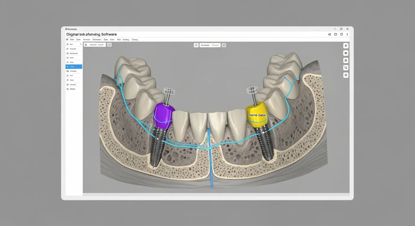
Millimeter-Precise Planning
Implant position, bone volume, and nerve pathways are made exactly visible before the procedure.
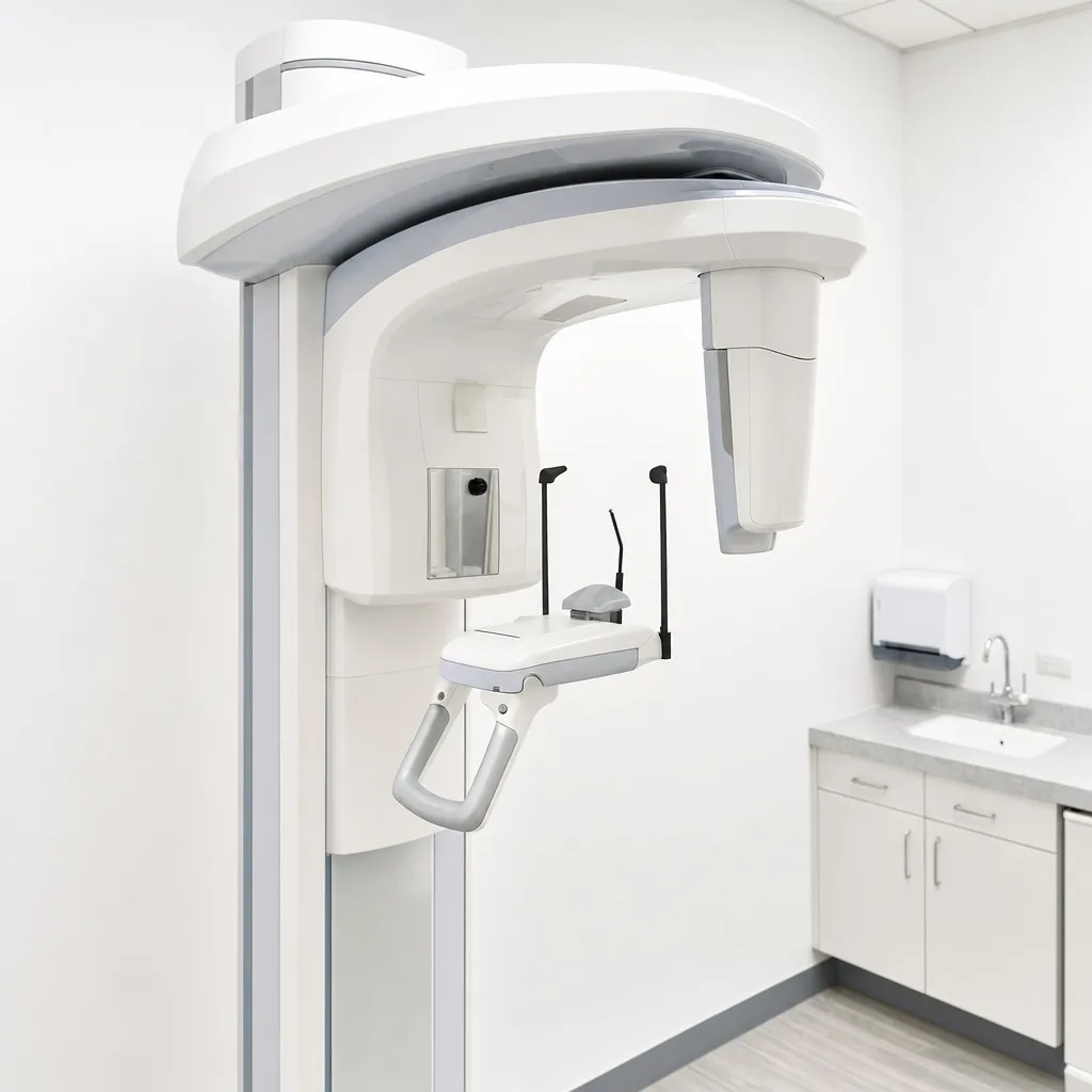
Precise planning begins with an exact image: with digital volume tomography, we display bony structures three-dimensionally the foundation for safe surgical planning.
A surgical procedure in the field of oral, maxillofacial, and facial surgery should always be based on comprehensive, detailed preoperative diagnostics. Nerves and other sensitive anatomical structures in the head and neck area lie particularly close together there.
Before every surgical procedure, all conceivable risks are carefully considered in order to avoid them from the outset wherever possible.
In addition to conventional two-dimensional X-rays (orthopantomogram), modern radiology today offers the possibility of computer-assisted 3D diagnostics and planning using digital volume tomography (DVT).
With razor-sharp 3D views, the bony structures of the upper and lower jaw, selected individual segments, or complex anatomical relationships can be displayed in detail for treatment planning that is precisely tailored to you.
Three reasons why we rely on three-dimensional imaging for more complex procedures.
Implant position, bone volume, and nerve pathways are made exactly visible before the procedure.
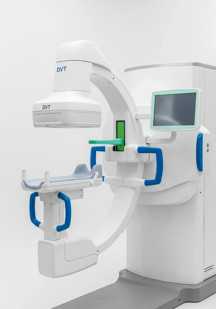
Sensitive structures such as nerves and the maxillary sinus are reliably displayed and taken into account during planning.
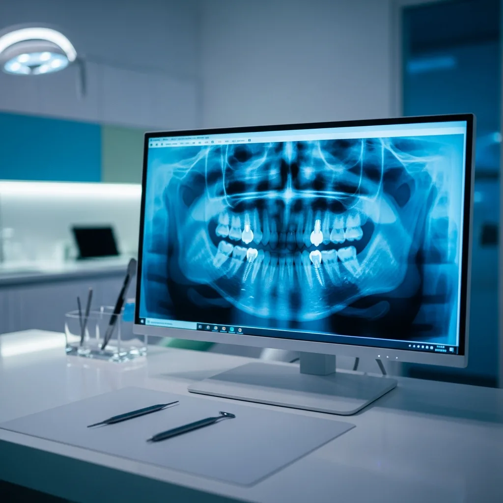
The 3D images can be shown directly on screen and reviewed together with you.
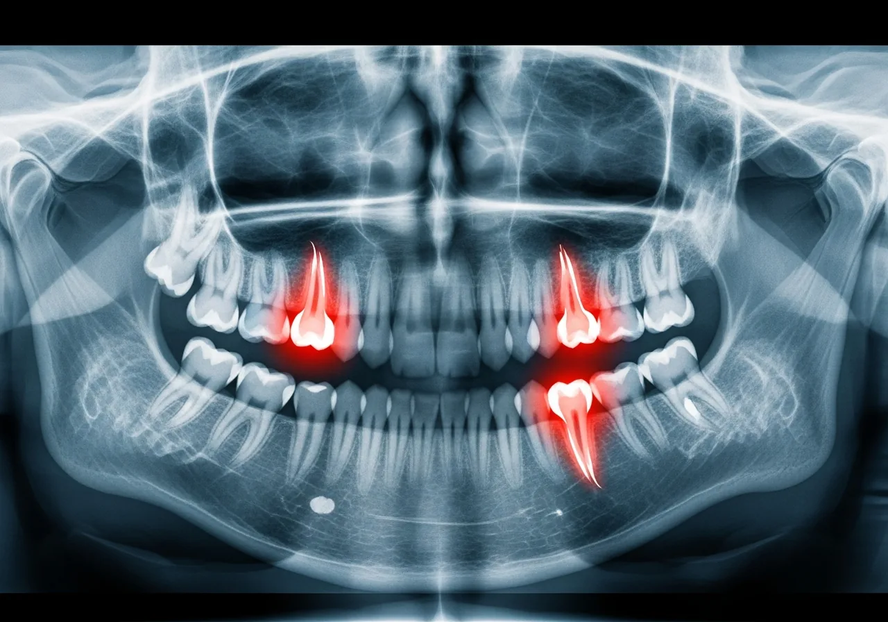
Placing dental implants or a surgical procedure in the jaw area requires careful planning. In addition to classic two-dimensional X-rays, we use computer-assisted 3D planning to predict the procedure as precisely as possible in advance.
This allows us to assess, even before treatment, which approach is gentlest for your individual situation for example, whether bone augmentation is necessary or an implant can be placed directly.